
FIGURE
Fig. 2
Fig. 2
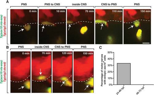
sox10+ peripheral cells migrate into the spinal cord during early nervous system development. A: Images from a 24 h time-lapse movie starting at 24 hpf of a Tg(sox10:nls-eos);Tg(olig2:dsred) embryo. Arrows denote a peripheral sox10+ cell that entered the spinal cord and then quickly exited. Dashed line denotes edge of spinal cord. B: 90-degree digital rotation of images from movie shown in A. C: Percentage of the number of nerves that had peripheral sox10+ cells enter the spinal cord and then exit (n = 9). Scale bar, 25 µm. |
Expression Data
Expression Detail
Antibody Labeling
Phenotype Data
Phenotype Detail
Acknowledgments
This image is the copyrighted work of the attributed author or publisher, and
ZFIN has permission only to display this image to its users.
Additional permissions should be obtained from the applicable author or publisher of the image.
Full text @ Glia