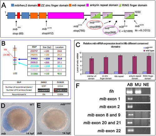
mib gene is deleted in mibnn2002 mutants. (A) Diagram of the mutations of different mib alleles. The translation of mib in mibnn2000 and mibnn2001 mutants stops at amino acids 5′ to ring finger domain 3 (RF3). (B) The linkage analysis result of mibnn2002 mutants. The upper part illustrates the locations of mib gene and the SSLP markers. The lower part demonstrates a close linkage of Z4662 and Z13620 to mib, with a recombination frequency of 0/97 and 2/214, respectively. (C) Real-time PCR results of mib expression in mibnn2002 homozygotes and WT embryos. The blue bars represent the expression level of mib in mibnn2002 homozygotes. The purple bars represent the expression level of mib in WT embryos. There is nearly no mib transcript detected in the mibnn2002 homozygotes. (D) WT embryos and (E) mibnn2002 mutants were stained with mib at 14 hpf, respectively. There is no mib mRNA detected in (E) mibnn2002 homozygous. (F) The results of mib genomic PCR were shown with fih (hif1an) as a positive control. AB represents AB WT samples; MU represents mibnn2002 homozygous samples; NE represents negative controls, where distilled water was used instead of genomic extracts as templates. The results showed that no mib genomic fragments are amplified.
|
