
Fig. 5
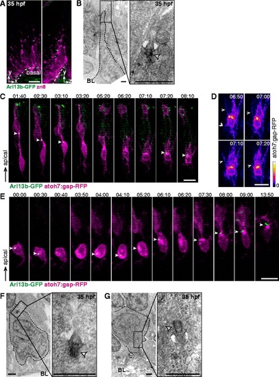
Upon completion of apical retraction, primary cilia may transiently disappear or lose their apical localization. a 3D maximum intensity projections of a region of the retina of a 35 hpf transgenic embryo expressing Arl13b-GFP and immunolabaled with zn8 antibody to identify RGCs. Primary cilia are observed colocalizing with the apical tip of cells finishing retraction. b TEM micrograph from a 35 hpf embryo showing a primary cilium at the tip of an apical process. The white arrowhead indicates the basal body. c-e Blastomeres from transgenic embryos (atoh7:gap-RFP/Arl13b-GFP) were transplanted into wild type hosts. The resulting embryos were imaged by time-lapse confocal microscopy from around 30 hpf onwards. Montages from 3D maximum intensity projections of the stacks are shown. (d) Detail of the cell shown in (c), where initial filopodial activity (arrowheads) previous to dendrite formation is observed; in order to highlight atoh7:gap-RFP signal intensity, a color ramp is used. The full white arrowheads indicate the presence and position of the primary cilium in c and e. f-g TEM micrographs from 35 hpf embryos, showing cells located in the basal region of the neuroepithelium (RGC neuroblasts) with primary cilia both in apical (f) and basal (g) positions. The white arrowhead indicates the basal body. BL: basal lamina. Scale bars: A, 10 µm; B, 1 µm; C-E, 10 µm; F-G, 1 µm |