
Fig. 8
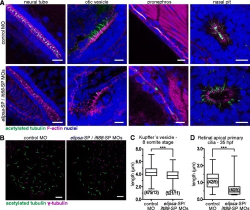
Effective reduction of primary cilia length in zebrafish embryos upon elipsa and ift88 knock-down. a Confocal images of different ciliated organs from 48 hpf embryos. Cilia were labeled with an anti-acetylated tubulin antibody and F-actin with TRITC-phalloidin. b Kupffer’s vesicle of eight-somite stage embryos, where basal bodies were labeled with an anti-γ-tubulin antibody. c Comparison of primary ciliary length in Kupffer’s vesicle. The experiments were performed twice, with similar results; only the results from one of the experiments are shown. d Comparison of the length of apical primary cilia in the retina of 35 hpf morphant and control embryos. Measurements were made on TEM micrographs. In C and D the numbers in brackets represent the number of cilia and embryos analyzed in each condition. (***) p < 0.001, Mann-Whitney test. Scale bars: A-B, 10 µm |