
Fig. 3
- ID
- ZDB-FIG-160505-25
- Publication
- Tan et al., 2016 - Stress from Nucleotide Depletion Activates the Transcriptional Regulator HEXIM1 to Suppress Melanoma
- Other Figures
- All Figure Page
- Back to All Figure Page
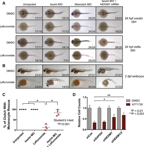
Knockdown of HEXIM1 Rescues Nucleotide Stress-Associated Neural Crest Ablation and Melanoma Suppression Phenotypes (A) Zebrafish embryos were injected with 8 ng hexim1 or control morpholino (MO) or a combination of hexim1 MO and 300 pg human HEXIM1 mRNA. Embryos were treated with DMSO or 6.5 µM lef at 50% epiboly and in situ hybridization was performed at 24 hr post-fertilization (hpf) for crestin or mitfa expression. Dorsal views of embryos are shown. Numbers indicate the number of embryos with the shown phenotype versus the total number of embryos in the clutch. (B) Embryos treated as in (A) were scored for melanocytes at 2 dpf. (C) Four clutches of embryos were analyzed for rescue of melanocytes under the conditions of uninjected, hexim1 MO injected, lef treatment, or hexim1 MO injected with lef treatment. Percentages of the number of embryos rescued in each clutch are plotted (mean ± SD). (D) Cell number of A375 cells treated with 25 µM A771726 in combination with siRNA pools for GAPDH, HEXIM1, and HEXIM2 relative to DMSO controls is shown (mean of three replicates ± SD). See also Figure S3. |
| Genes: | |
|---|---|
| Fish: | |
| Condition: | |
| Knockdown Reagent: | |
| Anatomical Term: | |
| Stage: | Prim-5 |
| Fish: | |
|---|---|
| Condition: | |
| Knockdown Reagent: | |
| Observed In: | |
| Stage Range: | Prim-5 to Long-pec |