
Fig. 3
- ID
- ZDB-FIG-160428-21
- Publication
- Jimenez et al., 2016 - Phenotypic chemical screening using zebrafish neural crest reporters identifies retinoid acid as an inhibitor of epithelial morphogenesis
- Other Figures
- All Figure Page
- Back to All Figure Page
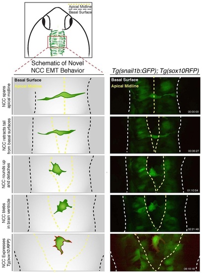
Dorsal midline neural epithelial progenitor cells display novel delamination behaviors of EMT. Schematic illustrating imaging area (top) and novel delamination behavior of a dorsal midline neuroepithelial progenitor (left); the schematic is derived from confocal time-lapse images of Tg(snai1b:GFP); Tg(sox10:RFP) embryos (right). Images show a GFP-positive cell initially spanning the dorsal midline before contracting apical attachments from each side of the neuroepithelium. Subsequently, GFP-positive cells detach and become rounded with filopodia and blebbing protrusions, and ultimately express sox10-mRFP at cell membranes. See also Movie 3. |