
FIGURE
Fig. 3
- ID
- ZDB-FIG-160421-7
- Publication
- Johansen et al., 2016 - Optical micromanipulation of nanoparticles and cells inside living zebrafish
- Other Figures
- All Figure Page
- Back to All Figure Page
Fig. 3
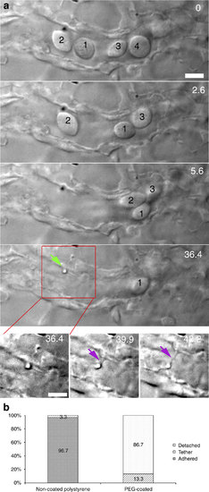
Multiple traps for in vivo nanotube formation in cell-free blood vessels. (a) In a smaller blood vessel several erythrocytes are cleared (0-36.4 s) and fenced off, after which an adhered particle was moved away from the endothelium, and a tethering nanotube was formed (36.4-42.2 s). Scale bar, 5 µm. (b) Quantification of the adhesion probability of naked and PEG-coated particles, classified as: detachable (white), strongly adhered (solid) and tethered (lines). Experiment was repeated at least 60 times. |
Expression Data
Expression Detail
Antibody Labeling
Phenotype Data
Phenotype Detail
Acknowledgments
This image is the copyrighted work of the attributed author or publisher, and
ZFIN has permission only to display this image to its users.
Additional permissions should be obtained from the applicable author or publisher of the image.
Full text @ Nat. Commun.