
Fig. 1
- ID
- ZDB-FIG-160421-5
- Publication
- Johansen et al., 2016 - Optical micromanipulation of nanoparticles and cells inside living zebrafish
- Other Figures
- All Figure Page
- Back to All Figure Page
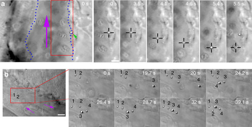
In vivo optical micromanipulation of microinjected particles. (a) A particle (green arrowhead) adhered to the endothelium of the caudal vein (indicated with blue dotted lines) is pulled away from the endothelium into the fast blood flow (purple arrow) using optical tweezers (black crosshairs). At time 5.5 s an erythrocyte is drawn into the trap. This replaces the particle in the trap which is subsequently pulled back towards the original adhesion point of the endothelium, presumably due to a connecting nanotube. Experiment repeated at least 80 times. (b) Four separate particles (numbered) are fished out of the blood flow and moved towards a sheltered region at the tip of the tail. Purple arrows indicate flow direction. Experiment was repeated at least 10 times. Scale bar, 5 µm. |