
Fig. S3
- ID
- ZDB-FIG-160415-17
- Publication
- Fernández-Murray et al., 2016 - Glycine and Folate Ameliorate Models of Congenital Sideroblastic Anemia
- Other Figures
- All Figure Page
- Back to All Figure Page
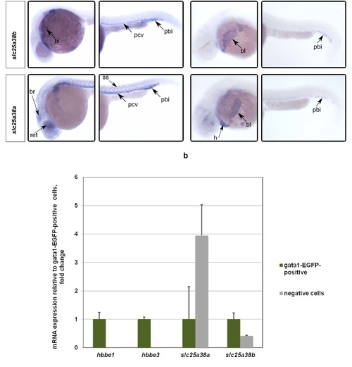
Whole-mount in situ hybridizations with probes for slc25a38a and slc25a38b at 24 and 34 hpf stages of development demonstrate predominant erythroid expression. To confirm preferential expression in red blood cells, EGFP-positive erythrocytes were isolated by FACS from gata1:EGFP (where erythroid lineage cells are labeled by EGFP) transgenic zebrafish embryos. Quantitative PCR assays for hbbe1, hbbe3, slc25a38a and slc25a38b were performed on cDNA made from FACS-sorted positive and negative cells. Gene expression qPCR values were normalized to 18S ribosomal RNA values. Relative expression values to EGFP-positive cells are presented. Error bars indicate standard errors from quantitative PCR experiments done on 3 separate FACS-sorted cell samples. *** p < 0.001, ** p < 0.01, * p < 0.05 for the t-test between expression values for the EGFP-positive and negative cells. |
| Genes: | |
|---|---|
| Fish: | |
| Anatomical Term: | |
| Stage: | Prim-5 |