
Fig. 2
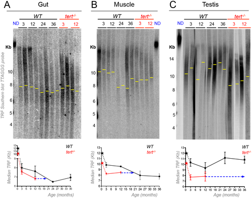
Telomeres shorten in WT gut and muscle, but not in testis, with aging reaching the length of tert-/- tissues. A-C) Representative images of telomere restriction fragment (TRF) analysis of genomic DNA by Southern Blot (random primer-labelled telomeric probe (TTAGGG)n 32P-dCTP) and quantifications of median telomere length. Black and red stars represent WT and tert-/- larvae telomeres, respectively, in quantifications of median TRF over time. WT telomeres shorten linearly with aging from 3 to 24 months, in A) gut (N = 3–4 per time point) and B) muscle (N = 3–4 per time point), stabilizing in later ages. C) No significant telomere shortening is detected in the testis (N = 5–6 per time point). Around 20 months of age WT telomeres reach the shorter length of 12 month-old tert-/- in the gut (graph in fig A–ca. 6.8 Kb) and muscle (graph in fig B–ca. 8.5 Kb) but not in testis (graph in fig C), indicated by blue arrow. N = 3–4 per time point for tert-/- gut and muscle, N = 4–6 per time point for tert-/- testis. TRF mean sizes were calculated as previously described [54]. Data are represented as mean +/- SEM. |
| Fish: | |
|---|---|
| Observed In: | |
| Stage: | Adult |