
Fig. 10
- ID
- ZDB-FIG-160226-9
- Publication
- Miyagi et al., 2016 - Characterization of the zebrafish cx36.7 gene promoter: Its regulation of cardiac-specific expression and skeletal muscle-specific repression
- Other Figures
- All Figure Page
- Back to All Figure Page
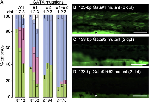
Mutational analysis of the 133-bp promoter. (A) Fertilized one-cell eggs were injected with the 133-bp promoter construct (WT) or its mutants with point mutations in either one or both of the GATA #1 and #2 elements. EGFP signals in the embryos were analyzed at 1, 2 and 3 dpf. The stacked bar charts represent the percentages of the following four groups of embryos: no EGFP expression (blue), cardiac-specific EGFP expression (green), EGFP expression in the heart and trunk muscle (red), and dead embryos (white). Data are expressed as the mean ± SEM of three independent experiments. (B–D) Representative images of the trunk region of 2-dpf embryos injected with the 133-bp promoter constructs having mutations in GATA #1 (B), GATA #2 (C), and both GATA #1 and GATA #2 (D). The asterisks indicate yolk sac autofluorescence. Bars, 0.2 mm. |
Reprinted from Gene, 577(2), Miyagi, H., Nag, K., Sultana, N., Munakata, K., Hirose, S., Nakamura, N., Characterization of the zebrafish cx36.7 gene promoter: Its regulation of cardiac-specific expression and skeletal muscle-specific repression, 265-74, Copyright (2016) with permission from Elsevier. Full text @ Gene