
Fig. 3
- ID
- ZDB-FIG-160225-40
- Publication
- Gokey et al., 2016 - A Kupffer's vesicle size threshold for robust left-right patterning of the zebrafish embryo
- Other Figures
- All Figure Page
- Back to All Figure Page
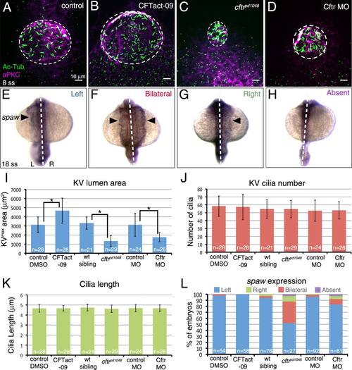
Role of Kupffer′s vesicle size in LR patterning. A–D: Modulating Cftr activity was used to alter KV lumen size at 8 ss. The Cftr activating drug CFTact-09 increased KV size (B), whereas a loss-of-function mutation (C) or morpholino (MO) interference (D) reduced KV size relative to controls (A). Dashed circles represent approximate KV lumen boundaries. E–H: Possible outcomes of RNA in situ hybridization analysis of the LR patterning marker spaw in lateral plate mesoderm (arrowhead) at 18 ss are normal left-sided (E), bilateral (F), reversed right-sided (G), or absent (H) expression. Dashed lines indicate the embryonic midline. L = left; R = right. I–K: Quantification of KV area (I), cilia number (J), and cilia length (K) in embryos with altered Cftr function and controls. L: Analysis of spaw expression in Cftr modulated embryos. Error bars represent one standard deviation. N = number of embryos analyzed. * indicates P < 0.05. |