
Fig. 1
- ID
- ZDB-FIG-160210-6
- Publication
- Dempsey et al., 2015 - Determination of the source of SHG verniers in zebrafish skeletal muscle
- Other Figures
- All Figure Page
- Back to All Figure Page
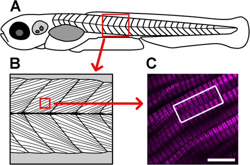
Verniers are regions of distorted endogenous muscle myosin SHG (magenta) that are visible deep within the somite. (A) This cartoon depicts a 5 dpf larva (not to scale). The red boxed area highlights a portion of the skeletal muscle compartment, which is illustrated in more detail in (B), showing the oriented arrays of muscle cells within the tissue. (C) In this image from a 5 dpf, laterally mounted, and fixed WT larva, clear distortions can be appreciated in the SHG signal within muscle cells (curvature toward the edges of the SHG bands in much of the image). The white boxed region draws attention to SHG verniers that appear to span across adjacent muscle cells, suggesting potential physical connections between cells across their membrane boundaries. Scale bar: 10 µm. |