
FIGURE
Fig. S1
- ID
- ZDB-FIG-160208-22
- Publication
- Sisson et al., 2015 - A role of glypican4 and wnt5b in chondrocyte stacking underlying craniofacial cartilage morphogenesis
- Other Figures
- All Figure Page
- Back to All Figure Page
Fig. S1
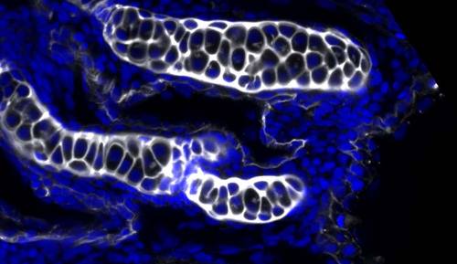
. Examples of wnt11 derived mutants. (A–B) Confocal images of a single focal plan of WGA staining (gray) of Wnt/PCP mutant craniofacial cartilage elements at 4 dpf. Nuclei were labeled with DAPI (blue). Scale bar = 20 µm. (A) A wnt11 embryo with a cyclopic index of 2 demonstrating while the cartilage elements are deformed, chondrocytes are still able to elongate. (B) Confocal images of a single focal plan of a wnt5b; wnt11 double mutant demonstrating the loss of chondrocyte elongation and stacking similar to the wnt5b single mutant. |
Expression Data
Expression Detail
Antibody Labeling
Phenotype Data
Phenotype Detail
Acknowledgments
This image is the copyrighted work of the attributed author or publisher, and
ZFIN has permission only to display this image to its users.
Additional permissions should be obtained from the applicable author or publisher of the image.
Reprinted from Mechanisms of Development, 138 Pt 3, Sisson, B.E., Dale, R.M., Mui, S.R., Topczewska, J.M., Topczewski, J., A role of glypican4 and wnt5b in chondrocyte stacking underlying craniofacial cartilage morphogenesis, 279-90, Copyright (2015) with permission from Elsevier. Full text @ Mech. Dev.