
Fig. 6
- ID
- ZDB-FIG-160208-21
- Publication
- Sisson et al., 2015 - A role of glypican4 and wnt5b in chondrocyte stacking underlying craniofacial cartilage morphogenesis
- Other Figures
- All Figure Page
- Back to All Figure Page
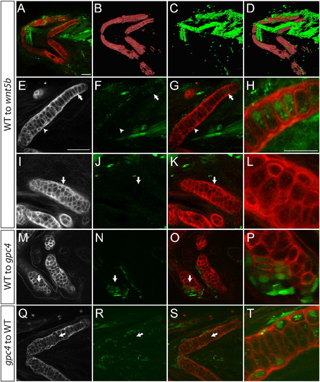
In craniofacial cartilage gpc4 acts autonomously. Transplantations to test for cell autonomy. (A–H) Transplanted wild type cells appear to rescue the wnt5b phenotype in the ceratohyal. (B–D) 3D reconstruction of panel A. (E–H) The ceratohyal with transplanted cells shown in A. Arrow indicates elongated transplanted cells surrounded by mesodermal transplanted cells. (H) Enlargement of area indicated in E–G by the arrow. (I–L) The certatohyal that is not transplanted with wild type cells in panel A retains its wnt5b phenotype. (I–K) Arrows indicate region of enlargement shown in L. (M–P) Wild type transplanted cells into a gpc4 Meckel′s cartilage are cell autonomous and elongate (arrow). (P) Enlargement of M–O at arrow. (Q–T) gpc4 transplanted cells to a wild type host are cell autonomous and are rounder than wild type cells. (T) Enlargement of L–N at arrow. (E–Q) Wheat Germ Agglutinin (WGA) staining of ceratohyal cartilage. (F, J, N and R) Transplanted cells marked by streptavidin. (A–G, I–K, M–O, Q–S) scale bars = 50 µm. (H, L, P, T) scale bar = 25 µm. WGA (red), transplanted cells marked by Alexa-Streptavidin (green). |
Reprinted from Mechanisms of Development, 138 Pt 3, Sisson, B.E., Dale, R.M., Mui, S.R., Topczewska, J.M., Topczewski, J., A role of glypican4 and wnt5b in chondrocyte stacking underlying craniofacial cartilage morphogenesis, 279-90, Copyright (2015) with permission from Elsevier. Full text @ Mech. Dev.