
FIGURE
Fig. S1
- ID
- ZDB-FIG-151214-49
- Publication
- Roxo-Rosa et al., 2015 - The zebrafish Kupffer's vesicle as a model system for the molecular mechanisms by which the lack of Polycystin-2 leads to stimulation of CFTR
- Other Figures
- All Figure Page
- Back to All Figure Page
Fig. S1
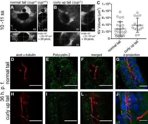
cup mutant KV volumes. Whole KV live-microscopy scans of 10–11 s.s. embryos from cup+/- ; foxj1a:GFP parents. The middle focal plan along the XY axis and the respective orthogonal views (along XZ and YZ axes) are shown for the most representative normal tail (cup+/-; cup+/+) (A), and curly-up tail (cup-/-) (B) embryos. KVvolume is indicated in µm3 and in picoL. Scale bars: 10 µm. (C) Estimated KV volumes (µm3) for normal tail (cup+/-; cup+/+) (n = 23) and curly-up tail (cup-/-) (n = 15) embryos. Average values and the respective s.d. are indicated. |
Expression Data
| Genes: | |
|---|---|
| Antibodies: | |
| Fish: | |
| Anatomical Terms: | |
| Stage Range: | 10-13 somites to Prim-25 |
Expression Detail
Antibody Labeling
Phenotype Data
| Fish: | |
|---|---|
| Observed In: | |
| Stage: | 10-13 somites |
Phenotype Detail
Acknowledgments
This image is the copyrighted work of the attributed author or publisher, and
ZFIN has permission only to display this image to its users.
Additional permissions should be obtained from the applicable author or publisher of the image.
Full text @ Biol. Open