|
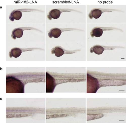
Analysis of miR-182 expression in 30 hpf embryos. (a-c) Whole-mount in situ hybridization of 30 hpf wild type embryos using miRCURY LNATM microRNA Detection Probes for miR-182 (miR-182-LNA, left panel) or a scrambled control (scrambled-LNA, middle panel). Right panel shows images of embryos incubated without LNA probe. (b) Higher magnification images of (a) showing the yolk sac and trunk region. (c) Higher magnification images showing soley the trunk region. miR-182 expression can be detected in eye lens, olfactory placode, optic tectum and notochord. Size bar corresponds to 250 µm in (a) and 125 µm in (b, c).
|