Fig. 7
- ID
- ZDB-FIG-151102-34
- Publication
- Ghaye et al., 2015 - Progenitor potential of nkx6.1-expressing cells throughout zebrafish life and during beta cell regeneration
- Other Figures
- All Figure Page
- Back to All Figure Page
|
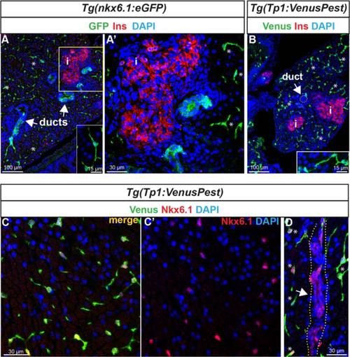
Expression of nkx6.1 persists in duct cells in the adult pancreas. a GFP and insulin (Ins, red) immunodetection on section through the pancreas of adult Tg(nkx6.1:eGFP) zebrafish. White arrows point to pancreatic ducts and asterisks show cells (presumably centroacinar/ terminal end duct cells (CACs) dispersed throughout the exocrine tissue). a′ Close-up of the islet highlighted with Ins. b Venus (green) and Ins (red) immunodetection in Tg(Tp1:VenusPest) showing the presence of Venus in CACs (asterisks) as previously reported 12], but not in duct cells within ductular structures (white arrow). Insets in (a) and (b) show isolated CACs. c–d Immunodetection of Venus (green) and of endogenous Nkx6.1 (red) in Tg(Tp1:VenusPest) revealing co-labeling of both markers in CACs (c, c′) while Nkx6.1 alone, but not Venus, labels the ducts. The white arrow in (d) points to a duct and asterisks indicate CACs. Dotted yellow lines delimit the duct. c′ Same as (c) showing Nkx6.1 (red) and 4′,6-diamidino-2-phenylindole (DAPI) only. i, islet |
| Genes: | |
|---|---|
| Antibody: | |
| Fish: | |
| Anatomical Terms: | |
| Stage: | Adult |

