
Fig. 1
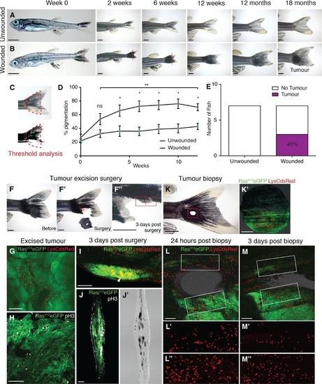
Tumours arise at sites of injury. A. Unwounded 4weekold kita:RasG12VeGFP juvenile fish followed over time. B. Juvenile kita:RasG12VeGFP fish wounded by tailfin resection (red line) every 2 weeks for 12 weeks. C. Pigmentation of the tail fin as quantified by threshold analysis of the tail area (indicated by red dotted line) at each time point. D. Graph illustrating the percent pigmentation over 12 weeks (mean ± SEM). E. Number of tail fin tumours developed in wounded versus unwounded fish (n = 7 fish in unwounded and wounded group, respectively). F–F′′ An adult kita:RasG12VeGFP fish with a large tail melanoma prior to surgery (F), immediately after (F′) and 3 days postsurgery (F′′); the surgery left a small section of melanoma remaining on the ventral tail fin (highlighted by the red box). G. Wholemount immunohistochemistry of the excised unwounded tumour stained for LysC (red) to reveal neutrophils. H. Immunohistochemistry of a section of the same excised tumour stained for phosphohistone H3 (pH3, white) to reveal the extent of proliferation. I. Wholemount immunohistochemistry of the tail with wounded tumour remnant 3 days postsurgery (green) illustrating accumulation of neutrophils (red) within the wounded tumour tissue (arrowhead). J, J′ A frozen section through the tail with wounded tumour remnant 3 days postsurgery (plane indicated by red dotted line in F′′) stained for phosphoH3 revealing how proliferating cells (white) appear to have increased in number in the remaining tumour. Equivalent brightfield section shown in (J′). K, K′ An adult kita:RasG12VeGFP; p53+/-; LysC:dsRed fish with an earlystage, flat, tail melanoma just postpunch biopsy at the tumour margin; punch biopsy tissue with green RasG12VeGFP tissue adjacent to healthy tissue is shown in (K′). L–L′′ Multichannel view of the repairing wound illustrating how, 24 h postwounding, neutrophils (red) have been drawn to both tumour and healthy tissue wound edge. (L) and (L′′) are magnifications of the wound edges of nontumour and tumour tissue, respectively. M–M′′ Multichannel view of the repairing wound at 3 days postwounding, demonstrating that neutrophils have largely resolved away from the wound site in healthy tissue (M′) but remain highly concentrated in wounded tumour tissue (M′′). |
| Fish: | |
|---|---|
| Condition: | |
| Observed In: | |
| Stage Range: | Days 30-44 to Adult |