
Fig. S5
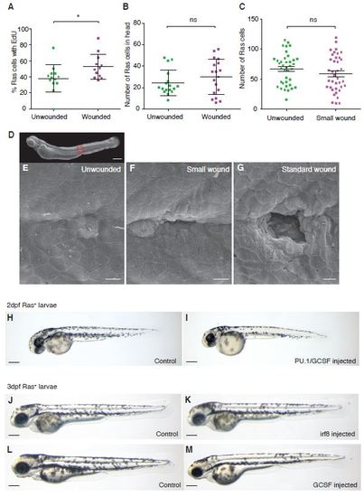
Only large wounds trigger proliferation in nearby pre-neoplastic cells. A. Graph showing the percentage of EdU positive pre-neoplastic cells in unwounded versus wounded larvae. B. The number of pre-neoplastic cells distant from the wound site (in the larval head) is unchanged between wounded and unwounded larvae. C. Graph to show that small wounds (as shown in the SEM image, G) do not lead to increased proliferation. D. shows a whole zebrafish larvae with a box in red to indicate where high magnification pictures were taken of an unwounded 3dpf larvae (E), a small wound (F) where ablation is restricted to just one epithelial cell (although adjacent cells may also be affected), and a standard sized wound (G) which causes broader tissue damage across a diameter of 5-7 cells (i.e. >35µm). H-M Brightfield images of Ras+ larvae injected with control morpholino (H, J and L) PU-1/GCSF morpholino (I) irf8 morpholino (K) and GCSF morpholino (M) at 2 or 3dpf (labelled). Scale bars represent 200µm (D and H - M) and 10µm (E-G). |