
Fig. 4
- ID
- ZDB-FIG-151002-50
- Publication
- Jank et al., 2015 - Tyrosine glycosylation of Rho by Yersinia toxin impairs blastomere cell behaviour in zebrafish embryos
- Other Figures
- All Figure Page
- Back to All Figure Page
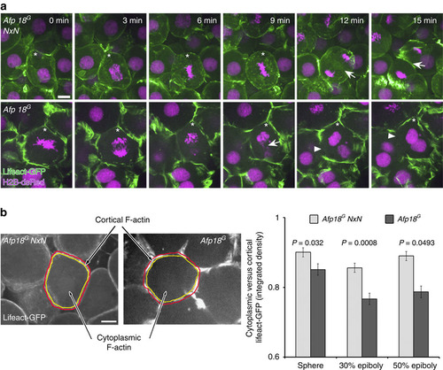
Afp18G blocks cytokinesis leading to multinucleated cells. (a) Cytoskeleton and cytokinesis analysed by time series of Afp18G NxN (upper row) or Afp18G (lower row) mRNA (0.5 pg per embryo each) injected embryos at dome stage (4.3 h.p.f.). Embryos were co-injected with mRNA encoding Lifeact-GFP (green) and H2B-dsRed (magenta; 100 pg per embryo each) labelling the F-actin cytoskeleton and the nuclei. Asterisk marks dividing blastomeres. Afp18G NxN and Afp18G mRNA-injected blastomeres complete microtubule mediated mitotic phases including chromosome segregation. Arrows indicate the actin ring contraction during cytokinesis, which is strongly affected in Afp18G-dividing blastomeres. Confocal z-stack projection of 10-µm depth; scale bar, 10 µm. (b) Quantification of cytoplasmic versus cortical Lifeact-GFP integrated epifluorescence signal density at indicated developmental stages. Confocal image of Afp18G NxN (left side) and Afp18G (right side) injected embryo showing an example blastomere with manually defined areas of cortical (region depicted in between red and yellow selection) and cytoplasmic (region outlined by yellow selection) F-actin and analysed using Fiji-ImageJ software measure function (sphere, n=12 each; 30% epiboly n=12 each; 50% epiboly n=12 each). Scale bar, 10 µm. Values are average±s.e.m. Statistical significance was evaluated using Mann–Whitney test. |