
Fig. 3
- ID
- ZDB-FIG-151002-49
- Publication
- Jank et al., 2015 - Tyrosine glycosylation of Rho by Yersinia toxin impairs blastomere cell behaviour in zebrafish embryos
- Other Figures
- All Figure Page
- Back to All Figure Page
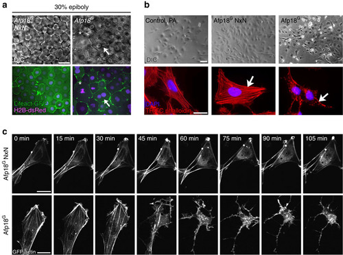
Afp18G affects the actin cytoskeleton. (a) Live images of Afp18G NxN or Afp18G mRNA (each 0.5 pg per embryo) injected embryos at 30% epiboly. Embryos are co-injected with mRNA encoding Lifeact-GFP (green) and H2B-dsRed (magenta; 100 pg per embryo each) labelling the F-actin cytoskeleton and the nuclei. Afp18G NxN embryos developed indistinguishable from WT embryos. Differential interference contrast (DIC) images of blastomeres are shown in the upper row, the corresponding confocal epifluorescence images below. Asterisk marks abnormally large blastomere; the arrow marks a blastomere with two nuclei. Single-plane image, scale bar, 20 µm. (b) Fluorescent micrographs of ZF4 cells treated with 6xHis-tagged Afp18G (right panel) or glycosyltransferase-deficient mutant Afp18G NxN (middle panel) proteins in combination with anthrax protective antigen (PA) as translocation system for His-tagged proteins. Top row shows phase-contrast images. Bottom row shows TRITC-phalloidin staining (red) of the actin cytoskeleton and a DAPI nuclei staining (magenta) of ZF4 cells fixed after 2 h. Arrows indicate regular stress fibres of F-actin in Afp18G NxN cells compared with disrupted F-actin fibres in Afp18G treated cells. Scale bar, 50 µm (top panel), 10 µm (bottom panel). (c) Time-lapse microscopic images of GFP-actin expressing HeLa cells intoxicated with Afp18G NxN (top row) and Afp18G (bottom row) as described in b. Scale bar, 10 µm. DAPI, 4′,6-diamidino-2-phenylindole. |