
Fig. 6
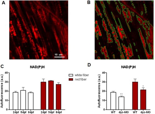
NAD(P)H signal is relatively stable during normal development but reduced in dystrophin morphants. (A) NAD(P)H signal of white skeletal muscle cells of zebrafish embryo at 5dpf. Scale bar,10µm. (B) Intensity-based segmentation of mitochondria. NAD(P)H signal was defined as mean intensity of mitochondrial regions. (C) NAD(P)H autofluorescence in the mitochondria of red (right) and white (left) skeletal muscle cells of wild-type embryos at 2, 5 and 8 dpf. No significant change of NAD(P)H was observed during development in either red or white fibers. N = 5–13 animals per group. (D) NAD(P)H signal was significantly decreased in both red and white fibers of dys-MO morphants (dys-MO) compared with wild-type (WT) controls. Data are expressed as the mean±SEM. *: p<0.05; **: p<0.01 versus wild-type group. |
| Fish: | |
|---|---|
| Knockdown Reagent: | |
| Observed In: | |
| Stage: | Day 5 |