
Fig. 2
- ID
- ZDB-FIG-150910-2
- Publication
- Fortuna et al., 2015 - Vascular Mural Cells Promote Noradrenergic Differentiation of Embryonic Sympathetic Neurons
- Other Figures
- All Figure Page
- Back to All Figure Page
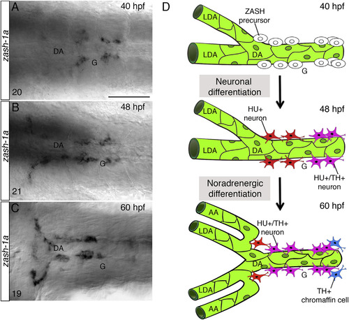
Early SN Precursors Develop Next to the DA (A–C) Whole-mount in situ hybridizations with antisense riboprobe specific for zash-1a RNA expression at the indicated time points. Images are ventral views (anterior is to the left) of the region between the LDA-DA connection (left) and the glomerular region (G) after dissection of the yolk sac. Zash-1a+ sympathetic precursors are present around the DA at 40 hpf (A). At 48 hpf (B) and 60 hpf (C), zash-1a expression expands to more anterior and posterior regions. The number of embryos observed is indicated at the bottom left. (D) Schematic representation of SN development. At 40 hpf, zash-1a+ sympathetic precursors differentiate next to the DA. At 48 hpf, they acquire HU expression and some of them become TH+. At 60 hpf, most of them have acquired TH expression. DA, dorsal aorta; LDA, lateral dorsal aorta; AA, aortic arch; G, glomerulus; hpf, hours post-fertilization. Scale bar, 75 µm (A–C). See also Figure S2. |