
Fig. S4
- ID
- ZDB-FIG-150910-11
- Publication
- Fortuna et al., 2015 - Vascular Mural Cells Promote Noradrenergic Differentiation of Embryonic Sympathetic Neurons
- Other Figures
- All Figure Page
- Back to All Figure Page
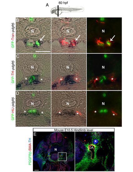
Pdgfr-β expression co-localizes with perivascular Transgelin positive cells, related to Figure 5. (A) Schematic representation of section plane in B-D. (B-D) Transversal section of 60 hpf Tg(kdrl:EGFP)la116 zebrafish embryos at the level of the vertical black bar in A. Embryos were analyzed for Pdgfr-β expression by in situ hybridization and immunostained with antibodies to detect GFP (vessels), Transgelin (Tran) (VMCs), Hu and TH. (B) Transgelin and pdgfr-β expression are co-localized in perivascular area (arrow). (C, D) pdgfr-β expressing cell do not co-localize with TH+ and HU+ cells (indicated by asterisk). (E) Transverse section of mouse embryo immunostained with antibodies against PDGFRβ (green), SMA (red) and TH (blue). Right panel shows magnification of the boxed area in left panel. PDGFRβ is expressed in VMCs, but not in SNs (asterisk). DA, dorsal aorta; N, notochord. Scale bars: 50 µm in B-E. |