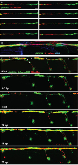
Regenerating axons followed Schwann cell processes. (A) Frames captured from time-lapse supplementary material Movie 4 of a Tg[gSAGFF202A;UAS:EGFP;SILL:mCherry] larva with severed axons, leaving a glial gap. Time elapsed from the first frame of the movie are, from left to right and top to bottom: 00:04, 00:20, 00:30, 00:40, 00:45, 00:46, 00:49, 00:52. Pictures were taken every 8min, starting 4h after axon severing. At 16hpt (00:45 time point), axons started to regrow when Schwann cells filled the gap. (B) An example of partially defasciculated axons at the site of injury. (C) A quadruple transgenic Tg[gSAGFF202A;UAS:EGFP;SILL:mCherry;Brn3c:mEGFP] larva starting at 6dpf and following 0.5hpt, 4hpt, 24hpt, 48hpt and 72hpt after axon severing and ablation of lateral Schwann cells. At 24hpt, regenerating axons re-innervated the neuromast with lateral Schwann cells, but failed to re-innervate neuromasts devoid of lateral Schwann cells even at 72hpt. Asterisks mark pigment cells. Arrows point to ablated lateral Schwann cells. Dash frame indicates the site of the cut. Scale bars: 25µm.
|
