
Fig. 4
- ID
- ZDB-FIG-150716-17
- Publication
- Xiao et al., 2015 - High-resolution live imaging reveals axon-glia interactions during peripheral nerve injury and repair in zebrafish
- Other Figures
- All Figure Page
- Back to All Figure Page
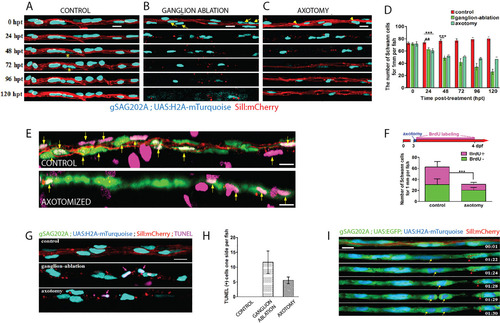
Differential responses of Schwann cells to acute and chronic denervation. (A-C) Tg[gSAGFF202A;UAS:H2A-mTurquoise;SILL:mCherry] larvae, starting at 5dpf and following 24hpt, 48hpt, 72hpt, 96hpt and 120hpt in the control (A), ganglion-ablated (B) and axon cuts (C) groups. Yellow asterisks mark the proliferative cells. Yellow arrows and arrowheads indicate the dead cells. (D) Quantification of Schwann cells, showing a significant reduction in the group with severed axons (n=6) and the ganglion-ablated group (n=8) compared with the control group (n=5) (**P<0.001, ***P<0.0001). (E) Control and axon-transected Tg[gSAGFF202A;UAS:EGFP;SILL:mCherry] larvae treated with BrdU. (F) The larvae were subject to axon severing at 3dpf in 10mM BrdU until 24hpt followed by fresh embryo medium. Fewer BrdU-positive Schwann cells were observed in the axon-transected (n=8) compared with the control (n=8) group (***P<0.0001, two-tailed t-test). (G) Examination of apoptotic Schwann cells by TUNEL assay. Pink arrows indicate colocalization of H2A-mTurquoise with TUNEL-positive cells. (H) Quantification of TUNEL-positive Schwann cells in the control group, the ganglion-ablation group and the axon-severed group. (I) Frames captured from supplementary material Movie 2 of a Tg[gSAGFF202A;UAS:EGFP;UAS:H2A-mTurquoise;SILL:mCherry] larva with severed axons. Numbers in the lower right corners denote time elapsed from the first frame of the movie. Red arrowheads mark axon debris that has been phagocytosed by the Schwann cells. At 10hpt (01:24 time point), Schwann cells started to divide (yellow arrow) when no intact axon exists. Error bars are+or±s.d. Scale bars: 10µm (A-C,E,G,I). |
| Genes: | |
|---|---|
| Fish: | |
| Conditions: | |
| Anatomical Terms: | |
| Stage Range: | Day 5 to Days 7-13 |