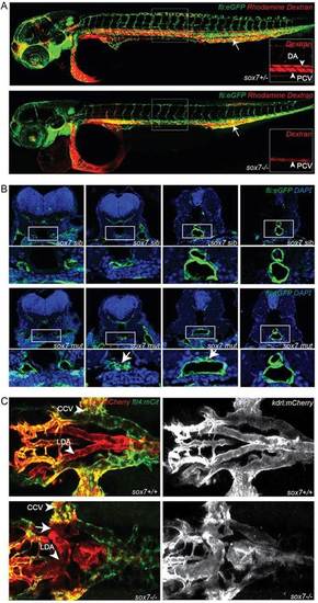
sox7 mutants show an altered morphology of the LDA while displaying normal DA-PCV segregation in the trunk. (A) Rhodamine-dextran angiograms (injected into the PCV at the position of the arrow) of sox7hu5626 fli1a:eGFP siblings (upper panel) and mutants (lower panel) at 2dpf. Insets depict the rhodamine-dextran channel of the boxed area with higher magnification highlighting the lack of dye uptake in the DA of sox7hu5626 mutants. (B) Transverse sections of fli1a:eGFP-positive sox7hu5626 sibling and mutant embryos at 2dpf, stained with anti-GFP (green) and DAPI (blue). In sox7hu5626 mutants, the aortic morphology is disturbed at the position of LDA/DA fusion (arrows) while being unaffected more anteriorly and posteriorly to this position. For relative positions of sections, see supplementary material Fig. S3. (C) Dorsal view of sox7hu5626 kdrl:mCherry;flt4:mCitrine sibling and mutant embryos at 2.5dpf. Right panel kdrl:mCherry channel only. Shunt formation (arrow) occurs in sox7hu5626 mutants at the position of LDA fusion. (L) DA, (lateral) dorsal aorta; PCV, posterior cardinal vein; CCV, common cardinal vein.
|
