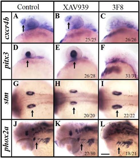
Modulating Wnt signaling around the shield stage alters the development of PPR-derived placodes. A–F: Marker gene expression changes in the rostral PPR-derived placodes of treated embryos. The XAV939 treatment (5–7 hpf) enlarges the olfactory cxcr4b expression (B) and lens pitx3 expression (E), compared with those in control embryos (A,D). The 3F8 treatment (5–7 hpf) leads to no olfactory cxcr4b (C) and lens pitx3 expression (F). G–L: Changes in the caudal PPR-derived placodal marker gene expression in treated embryos. The XAV939 treatment (5–7 hpf) reduces the otic stm (H) or does not change the epibranchial phox2a expression (K), compared with those of controls (G,J). The 3F8 treatment (5–7 hpf) results in larger otic stm (I) and stronger epibranchial phox2a expression (L). Arrows indicate the corresponding organ or organ primordium of each embryo. A–F, J–L: Side views with anterior to the left; G–I: Dorsal views with anterior to the left. A–I: 24 hpf; J–L: 48 hpf. Scale bar = 100 µm.
|
