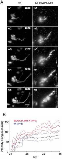
(A) Living isl-1 embryos were imaged by digital scanned laser light sheet microscopy from 24–36 hpf at 16min intervals. For analysis the images were deconvolved and the stacks were compressed into maximum projections. Representative images from control and MDGA2A morpholino treated embryos are shown. During the experimental period trigeminal neurons send out axons along a well-defined path, with no movement of the trigeminal neurons (w1–w5). In MDGA2A knockdown animals the compactness of the trigeminal cell cluster is impaired. Trigeminal neurons migrate along their axon bundles leaving their place of origin (bright dots along the axon bundle; m1–m5). (B) To quantify this migration phenotype, the time course of the fluorescence intensity along the trigeminal axon bundle was quantified in 8 control embryos and 5 MDGA2A morpholino treated embryos. Shown are the mean and bootstrap confidence intervals (as dotted lines, α = 0.05). Note that the fluorescence intensity along the trigeminal nerve in MDGA2A knockdown animals increases significantly past 30 hpf, representing the aberrant migration of trigeminal neurons. For more details see supplementary material Fig. S3A–C for individual intensity traces. The timestamp is hours post fertilization (h: min), N is the number of analyzed embryos, the scale bar is 20µm.
|
