
Fig. 1
- ID
- ZDB-FIG-150415-3
- Publication
- Ingold et al., 2015 - Proper migration and axon outgrowth of zebrafish cranial motoneuron subpopulations require the cell adhesion molecule MDGA2A
- Other Figures
- All Figure Page
- Back to All Figure Page
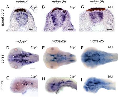
(A-C) Cross-section through larval zebrafish spinal cords. MDGA1 and MDGA2B staining can be observed at regions where dorsal commissural interneurons are located (white arrow). Additional staining for MDGA2A and MDGA2B can be observed in pools of intermediate and ventral interneuron. MDGA1 positive cells can be found in a narrow band of mediolateral located interneurons. Scale bars represent 25µm. (D–F) Dorsal and (G–I) lateral view of MDGA whole mount in situ hybridizations of 2dpf larval zebrafish. MDGA1 riboprobes label the ventral thalamus (asterisks in D,G) and the hypothalamus (arrow in D,G). Three well discernible cell clusters in the peripheral nervous system, namely the anterior and posterior lateral line ganglia as well as cells associated with the otic placode such as cells of the statoacoustic ganglion (gVIII) also prominently express MDGA1 (arrowheads in D,G). MDGA2A transcripts can be found in the motoneurons of the oculomotor (nIII) and trochlear (nIV) nerve in the midbrain (asterisks in E,H), as well as branchiomotoneurons of the trigeminal, facial and vagal nerve in the hindbrain (E,H, nV/nVII/nX). MDGA2B is expressed in the telencephalon, the ventral thalamus, the tegmentum, the hypothalamus and at low levels also in subpopulation (nVII and nX) of cranial motoneurons (F,I). Scale bars in D equals 100µm. |