
Fig. S2
- ID
- ZDB-FIG-150408-37
- Publication
- Shiau et al., 2015 - Differential Requirement for irf8 in Formation of Embryonic and Adult Macrophages in Zebrafish
- Other Figures
- All Figure Page
- Back to All Figure Page
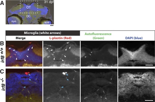
Thin brain sections reveal many microglial cells in wildtype but not in irf8 mutants, which appear to have only macrophages at 31 dpf. (A) The midbrain optic tectum, where many microglia normally reside, was the region of analysis. (B) Higher magnification of the brain region indicated in dotted box in A. 12–14 um cryosections were taken from wildtype irf8+/+ and heterozygous irf8st95/+ fish (n = 4) to compare with irf8st95/st9 mutants (n = 3) at 31 dpf. Immunostaining for L-plasin (with DAPI as a counterstain) was used to identify macrophages in relation to all cell bodies in the sections. Microglia were identified by their L-plastin expression, elaborate morphology with fine processes, and location in the parenchyma (white arrows). Cells in or adjacent to the interstitial space, vasculature, and ventricular zone were likely macrophages. Autofluorescence in the green channel helped to define the vasculature and distinguish actual L-plastin signal from vasculature autofluorescence in the red channel. irf8 mutants had a few L-plastin positive cells in the interstitial region and near the ventricular zone that lack fine processes (blue arrows), suggesting that these cells were not microglia. OT, midbrain optic tectum; DC, diencephalon. All scale bars are 50 um. |