
FIGURE
Fig. 7
- ID
- ZDB-FIG-150401-4
- Publication
- Roy et al., 2015 - NMDA receptors on zebrafish Mauthner cells require CaMKII-α for normal development
- Other Figures
- All Figure Page
- Back to All Figure Page
Fig. 7
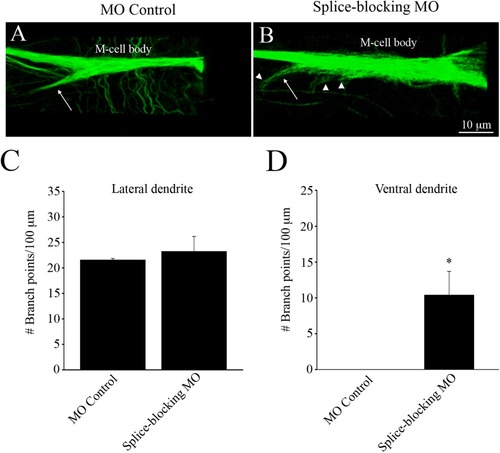
Immunohistochemical images of the M-cell labeled with anti-3A10, from control-injected and splice-blocking morphants. (A) The M-cell of MO control-injected fish. Arrow points to the ventral dendrite. (B) The M-cell of a splice-blocked embryo. Arrow points to the ventral dendrite. Arrowheads point to dendritic arborizations present on the cell body and ventral dendrite. Quantification of the number of branch points per 100 µm, emanating from the lateral dendrites (C) and the ventral dendrites (D). * Significantly different from MO controls, p < 0.05. |
Expression Data
| Antibody: | |
|---|---|
| Fish: | |
| Knockdown Reagent: | |
| Anatomical Terms: | |
| Stage: | Long-pec |
Expression Detail
Antibody Labeling
Phenotype Data
| Fish: | |
|---|---|
| Knockdown Reagent: | |
| Observed In: | |
| Stage: | Long-pec |
Phenotype Detail
Acknowledgments
This image is the copyrighted work of the attributed author or publisher, and
ZFIN has permission only to display this image to its users.
Additional permissions should be obtained from the applicable author or publisher of the image.
Full text @ Dev. Neurobiol.