
Fig. 9
- ID
- ZDB-FIG-150312-46
- Publication
- Feng et al., 2014 - Negative feedback regulation of Wnt signaling via N-linked fucosylation in zebrafish
- Other Figures
- All Figure Page
- Back to All Figure Page
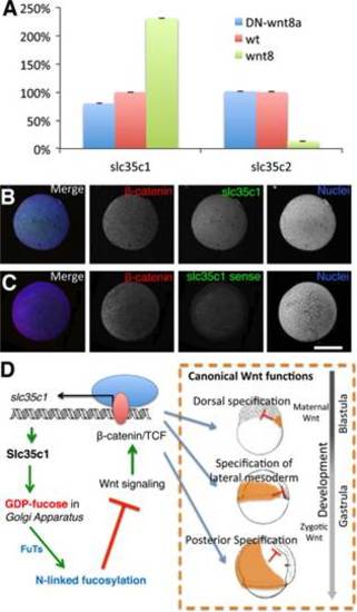
The interplay of Wnt8a signaling, slc35c1-mediated GDP-Fuc transport and N-linked fucosylation. (A) qPCR results show the relative expression of slc35c1 and slc35c2 in zebrafish embryos treated with wnt8a or DN-wnt8a mRNA and untreated control sphere stage (4hpf) embryos. All results are normalized to odc1. Expression in WT is defined as 100%. (B-C) Confocal images of slc35c1 fluorescent in situ show positive correlation between slc35c1 expression and β-catenin. Red: β-catenin protein staining. Green: in situ hybridization using slc35C1 antisense probe in (B) or slc35c1 sense probe in (C). Blue: nuclei. (D) A schematic model depicts the relationship between Slc35c1 and Wnt8a. Wnt8a activated canonical Wnt signaling enhances slc35c1 expression. Increased slc35c1 enhances N-linked fucosylation, which inhibits Wnt/β-catenin/TCF signaling during both blastula and gastrula stages. Therefore, these components comprise a negative regulatory loop to control Wnt signaling through N-fucosylation. Note: Bar=300 µm. |
| Gene: | |
|---|---|
| Antibody: | |
| Fish: | |
| Anatomical Terms: | |
| Stage: | Shield |
Reprinted from Developmental Biology, 395(2), Feng, L., Jiang, H., Wu, P., Marlow, F.L., Negative feedback regulation of Wnt signaling via N-linked fucosylation in zebrafish, 268-86, Copyright (2014) with permission from Elsevier. Full text @ Dev. Biol.