|
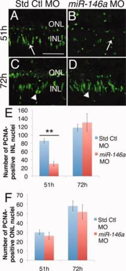
Knockdown of miR-146a reduces proliferation at 51h. Adult albino zebrafish were intravitreally injected and electroporated with either standard control or miR-146a morpholino prior to the start of light damage. A,B: After 51h of constant light, doublet PCNA-positive INL cells are present in standard control morphants (arrow), but mainly single PCNA-positive INL cells are present in miR-146a morphants (arrow). C,D: By 72h, both standard control and miR-146a morphants contained columns of INL proliferating nuclei (arrowheads). E,F: miR-146a knockdown significantly reduced the number of proliferating nuclei at 51h, but not at 72h. There was not a significant difference in the number of PCNA-positive INL cells at either 51h or 72h between the miR-146a and standard control morphant retinas. INL, inner nuclear layer; ONL, outer nuclear layer. Scale bar in = 50 µm and applies to B–D. Data represent mean ± s.e.m. **P< 0.01 using two-way ANOVA with Tukey′s post-hoc test, n=6.
|