
Fig. 4
- ID
- ZDB-FIG-150127-13
- Publication
- Hüsken et al., 2014 - Tcf7l2 Is Required for Left-Right Asymmetric Differentiation of Habenular Neurons
- Other Figures
- All Figure Page
- Back to All Figure Page
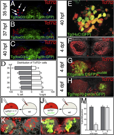
Tcf7l2 Is Expressed in Both Left- and Right-Sided dHbl and dHbm Neurons and Functions Cell/Lineage Autonomously (A–C, E–H, K, and L) Dorsal views with anterior to the top of the epithalamus at ages indicated and at 54 hpf (K and L). (A–D) Numbers of Tcf7l2 expressing cells (arrows in A) in Tg(foxD3:GFP); Tg(flh:eGFP) transgenic embryos are asymmetrically distributed in 35 and 37 hpf embryos (A and B) but are symmetric by 40 hpf (C and graph in D). Tcf7l2 expressing cells ventral to the habenulae are pseudocolored in blue. (E) High magnification of the left habenula stained for Tcf7l2 (red) and HuC:GFP (green). (F–H) Tcf7l2 expression (red) in wild-type (F), Et(gata2a:EGFP)pku588 (G), and Tg(hsp70-brn3a:GFP) (H) embryos colabeled with Sytox orange (gray nuclei). (I and J) Schematics of the experiment. RFP+ wild-type (I) or tcf7l2 mutant (J) cells carrying the pku588 transgene were transplanted into nontransgenic hosts in a region of the blastula from which cells later contribute to the habenulae. (K and L) Examples of transplanted control Et(gata2a:EGFP)pku588 × tcf7I2+/ or tcf7I2+/+ (K) and Et(gata2a:EGFP)pku588 × tcf7I2/ (L) cells (donor cells are red) in wild-type habenulae (nuclei labeled gray with DAPI). (M) dHb neurons from mutant donors expressing the pku588 transgene when incorporated into either the left or right habenula. Error bars represent the SD of the cell proportions. See also Figure S4 and Table S2. |
| Genes: | |
|---|---|
| Antibody: | |
| Fish: | |
| Anatomical Terms: | |
| Stage Range: | Prim-15 to Day 4 |