
Fig. 2
- ID
- ZDB-FIG-141028-2
- Publication
- Mich et al., 2014 - In vivo imaging of hedgehog pathway activation with a nuclear fluorescent reporter
- Other Figures
- All Figure Page
- Back to All Figure Page
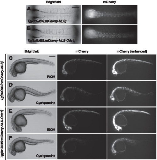
Tg(8xGliBS:mCherry-NLS-Odc1) zebrafish exhibit enhanced reporter turnover. (A–B) Brightfield and fluorescence images of 10-somite (~14 hpf) Tg(8xGliBS:mCherry-NLS) and Tg(8xGliBS:mCherry-NLS-Odc1) embryos. (C–F) The transgenic lines at 30 hpf after the addition of cyclopamine or vehicle control at the 10-somite stage. The embryos were concurrently treated with phenylthiourea (0.003%, w/v) to block pigmentation. Brightness-enhanced fluorescence images are also shown to more clearly depict mCherry-positive cells in cyclopamine-treated embryos. The zebrafish embryos in (A) and (B) are the same animals shown in (D) and (F), respectively. Embryo orientations: A–B, dorsal view and anterior left; C–F, lateral view and anterior left. Scale bars: A–B, 100 μm; C–F, 200 μm. |