
Fig. 5
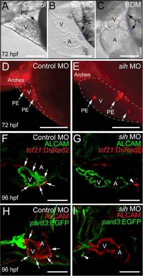
Heartbeat is not required for PE cluster formation, but is necessary for epicardium development. Lateral views of zebrafish hearts with anterior to the left. (A-C) Brightfield micrographs showing hearts from control (A), sih MO (B), and BDM-treated (C) fish at 72 hpf. The PE clusters are pseudo colored purple and indicated by arrows (n = 10 per group). (D and E) Epifluorescence images showing hearts from control (D) and sih MO-treated (E) fish at 72 hpf, using the tcf21:DsRed2 reporter to reveal PE clusters (arrows) (n = 15 per group). The pericardial space is outlined with a dashed line. (F-I) Confocal images of embryos treated with control and sih-MO collected at 96 hpf (n = 12 per group). (F and G)tcf21:DsRed2 is red and ALCAM is green. (H and I)pard3:EGFP is green and ALCAM is red. Arrows indicate epicardial cells developing across the ventricle. Arrows indicate small PE clusters expressing tcf21 or pard3. For all panels, V is ventricle; A, Atrium; BA, bulbus arteriosus. Scale bars = 50 microns. |