FIGURE
            Fig. 4
                
                    
                        Fig. 4


| 
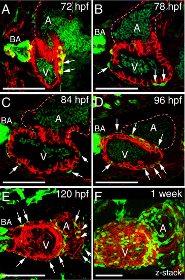
 Normal progression of epicardium formation. Ventral views of zebrafish hearts. (A-F) Epicardial cells are marked with pard3 (EGFP; green) and cardiomyocytes are marked with ALCAM (red). Confocal images from 72-120 hpf are optical slices showing progressive epicardium coverage (white arrows) proceeding across the ventricle (V) and then onto the atrium (A) at 120 hpf. The z-series at one-week shows epicardial cells on the ventricle and atrium, however epicardium coverage is not complete. For all panels, with anterior to the left and BA is bulbus arteriosus. Scale bars = 50 microns. For each time point n = 5. | 
                
                    
                        Expression Data


                        Expression Detail


                        Antibody Labeling


                        Phenotype Data


                        Phenotype Detail


                        Acknowledgments


This image is the copyrighted work of the attributed author or publisher, and
ZFIN has permission only to display this image to its users.
Additional permissions should be obtained from the applicable author or publisher of the image.
      Full text @ BMC Dev. Biol.


        