
Fig. 5
- ID
- ZDB-FIG-140612-5
- Publication
- Piotrowski et al., 2014 - The development of lateral line placodes: taking a broader view
- Other Figures
- All Figure Page
- Back to All Figure Page
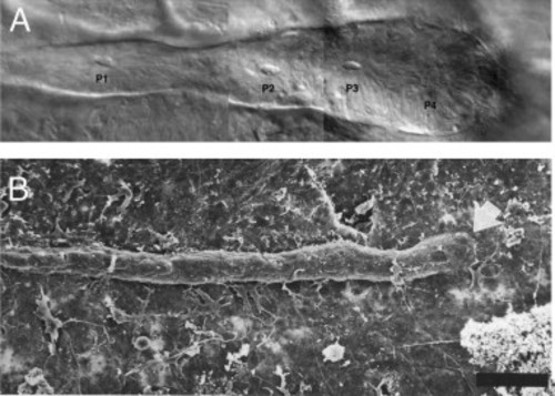
Posterior line placodes vary in shape. (A) Nomarski brightfield image of a posterior lateral line primordium in a blind Mexican tetra (blind cavefish, Astyanax fasciatus). The primordium is club-shaped and sensory organs begin to form within the migrating primordium (P1–P3) (reproduced from Sapède et al., 2002 with permission from Development). (B) Scanning electron micrograph of the posterior lateral line placode in an axolotl larva (Ambystoma mexicanum). The placode is much thinner than in teleosts. Image reproduced with permission from the International Journal of Developmental Biology ( Smith, 1996). Scale bar 100 µm. |
Reprinted from Developmental Biology, 389, Piotrowski, T., Baker, C.V., The development of lateral line placodes: taking a broader view, 68-81, Copyright (2014) with permission from Elsevier. Full text @ Dev. Biol.