
Fig. 2
- ID
- ZDB-FIG-140612-3
- Publication
- Piotrowski et al., 2014 - The development of lateral line placodes: taking a broader view
- Other Figures
- All Figure Page
- Back to All Figure Page
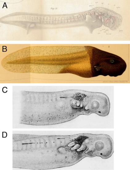
In the majority of non-cartilaginous vertebrates, the posterior lateral line placodes migrate and deposit lateral line sense organs. (A) The posterior lateral line placode often splits into a dorsal and a ventral component, as seen in the mudpuppy Necturus (an aquatic salamander) (reproduced from Platt, 1896). (B) Fusion of the trunk of a lightly pigmented frog embryo (Rana palustris) with the head of a darkly pigmented embryo (Rana sylvatica) demonstrated that pigmented lateral line placodes from the head migrate onto the trunk where they deposit sense organs ( Harrison, 1904). (C,D) Stone (1933) transplanted pieces of Nile blue-stained donor ectoderm containing the posterior lateral line placode onto an unstained spotted salamander (Ambystoma maculatum; Ambystoma punctatum) embryo. (C) 2 days post-transplantation the stained club-shaped placode had started to migrate. (D) By 3 days post-transplantation, the placodes had migrated further and deposited sense organs |
Reprinted from Developmental Biology, 389, Piotrowski, T., Baker, C.V., The development of lateral line placodes: taking a broader view, 68-81, Copyright (2014) with permission from Elsevier. Full text @ Dev. Biol.