
Fig. 3
- ID
- ZDB-FIG-140612-4
- Publication
- Piotrowski et al., 2014 - The development of lateral line placodes: taking a broader view
- Other Figures
- All Figure Page
- Back to All Figure Page
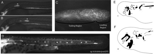
The posterior lateral line placode migrates; other lateral line placodes elongate to form sensory ridges. (A) At 24 hpf the posterior lateral line primordium (asterisk) in a zebrafish embryo has fully migrated onto the somites from its origin near the otic vesicle (OV). (B) The primordium has recently deposited the first proneuromast. (C) Higher magnification of the primordium in (A). Rosettes begin to form a short distance away from the leading region within the migrating primordium. (D) The post-embryonic lateral line is composed of 4–6 proneuromasts in a row along the horizontal myoseptum. Panels A–D are reproduced from Aman and Piotrowski (2011) with permission from Cell Adhesion and Migration. (E,F) Camera lucida drawings of flat mounts of the head ectoderm of axolotl embryos at (E) stage 25 and (F) stage 37, showing lateral line placodes in black and other placodes in gray. Panels E and F reproduced from Northcutt et al. (1994) with permission from Wiley and Sons. Abbreviations: ad, anterodorsal lateral line placode; av, anterodorsal lateral line placode; lp, lens placode; mid, middle lateral line placode; ol, olfactory placode; otv, otic vesicle; pld, dorsal subdivision of posterior lateral line placode; plm, main subdivision of posterior lateral line placode; plv, ventral subdivision of subdivision of posterior lateral line placode; st, supratemporal lateral line placode. |
Reprinted from Developmental Biology, 389, Piotrowski, T., Baker, C.V., The development of lateral line placodes: taking a broader view, 68-81, Copyright (2014) with permission from Elsevier. Full text @ Dev. Biol.