
Fig. 6
- ID
- ZDB-FIG-140523-34
- Publication
- Moya-Díaz et al., 2014 - Electroablation: a method for neurectomy and localized tissue injury
- Other Figures
- All Figure Page
- Back to All Figure Page
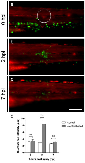
Neuromast electroablation induces an inflammatory response. Compound transgenic Tg(cxcr4b:mRFP; mpx:GFP) fish harboring neutrophils expressing green fluorescent protein and neuromasts labeled in red were used to study neutrophil inflammation induced by neuromast electroablation. Two 2 second 8 μA pulses were applied to induce damage to the pLL neuromast. (a-c) The trunk of a larva subjected to neuromast electroablation is shown with anterior to the left. (a) Immediately after electroablation, most neutrophils are present in the caudal hematopoietic tissue (CHT). (b) At 2 hpi, a large number of neutrophils have specifically migrated to the site of damage. (c) By 7 hpi, the number of neutrophils at the site of electroablation has diminished, suggesting resolution of inflammation is taking place. (d) Quantification of mean fluorescence in a 50 μm radius around the site of electroablation (panel a, dotted circle) as a measure of neutrophil recruitment. Data are presented as average ± SEM from 12 larvae per condition and two independent experiments. Comparisons were performed by using a repeated measurement two-way ANOVA, with Bonferroni’s post test. ***, p < 0.001; ns, p > 0.05. Scale bar (a-b) 100 μm. hpi, hours post-injury; a. u., arbitrary units. |