
Fig. 2
- ID
- ZDB-FIG-140523-30
- Publication
- Moya-Díaz et al., 2014 - Electroablation: a method for neurectomy and localized tissue injury
- Other Figures
- All Figure Page
- Back to All Figure Page
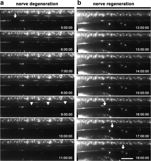
Neurectomy, degeneration and regeneration of the posterior lateral line (pLL) nerve. (a) Degeneration of a neurectomized pLL nerve after electrical injury. Transgenic TgBAC(neurod:EGFP) larvae with a labeled pLL nerve were neurectomized by applying a 17 μA pulse of current for 1.5 seconds. At 5 hpi a neurectomized larva was mounted in agarose for time-lapse imaging for 7 hours (anterior to the left). Fragmentation of the detached nerve fragment proceeds by a breakdown of the axons into many small sections simultaneously throughout the axotomized nerve (arrowheads). Note the intact contralateral pLL nerve which is visible (above the electroablated nerve, slightly out of focus) in all the images. (b) Regeneration of an axotomized pLL nerve. Transgenic TgBAC(neurod:EGFP) larvae was treated and imaged as before. In this case, the neurectomized larva was mounted for imaging for 6 hours starting at 12 hpi to examine regeneration of the pLL nerve. Note the progressive regeneration of the pLL nerve by elongation of the remaining axon stumps (arrows) as degeneration of the distal part of the axotomized pLL nerve has concluded. Scale bar, 50 μm; times in hh:mm:ss. |