
Fig. 4
- ID
- ZDB-FIG-140523-32
- Publication
- Moya-Díaz et al., 2014 - Electroablation: a method for neurectomy and localized tissue injury
- Other Figures
- All Figure Page
- Back to All Figure Page
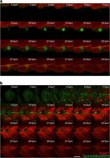
Extent of damage inflicted to tissues by neuromast electroablation. Transgenic Tg(-8.0cldnb:lynEGFP) fish, which express EGFP in lateral line cells and epithelial cells of the skin, were stained with BODIPY. Stained larvae were then mounted in agarose and subjected to electroablation of the L3 neuromast. Confocal images of electroablated and uninjured control larvae were acquired 20 minutes after injury with 1.5 μm of separation between z-axis optical slices. (a) Sequential z-axis confocal images of an uninjured larva are shown. The intact neuromast (green) exhibits a rosette-like structure and BODIPY-TR (red) allows visualization of underlying muscular tissue. (b) Images acquired as in (a) of a larva with an electroablated neuromast showing local loss of cells in the skin, disconnection of interneuromastic cells, destruction of neuromast cells and complete loss of the rosette-like structure. BODIPY-TR staining also shows a gap in muscular tissue to a depth of 22.5 μm from the skin surface; deeper sections appear unperturbed. Values indicate distance froom skin surface towards the inside of the larva in μm. Scale bar, 50 μm. |