
Fig. S3
- ID
- ZDB-FIG-140416-28
- Publication
- Elks et al., 2013 - Hypoxia inducible factor signaling modulates susceptibility to mycobacterial infection via a nitric oxide dependent mechanism
- Other Figures
- All Figure Page
- Back to All Figure Page
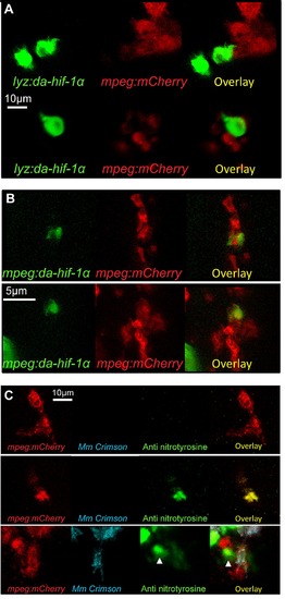
Leukocyte cell-type specific expression of stabilized Hif-1α and macrophage labeling with anti-nitrotyrosine. (A) Confocal photomicrographs of Tg(lyz:da-hif-1αb:ires-nlsegfp) (lyz:da-hif-1α) injected mpeg1:mCherry embryos at 2 dpf. IRES-nlsGFP is expressed in cells in the caudal haematopoetic tissue associated with leukocytes and mpeg1 positive macrophages, but is not present within the same cell. (B) Confocal photomicrographs of Tg(mpeg1:da-hif-1αb:ires-nlsegfp) (mpeg:da-hif-1α) injected Tg(mpeg1:mCherryF)ump2 line (mpeg:mCherry) at 2 dpf. IRES-nlsGFP is found expressed in the same cells as mpeg:mCherry indicating macrophage expression. (C) Confocal micrographs showing anti nitrotyrosine staining in macrophages in the mpeg:mCherry at 2 dpf. Upper panels show a nitrotyrosine negative macrophage, which is representative of the majority of the macrophage population. Middle panels show a nitrotyrosine positive macrophage in the absence of infection. Lower panels show a nitrotyrosine positive macrophage in the presence of infection. In both the absence and presence of infection nitrotyrosine positive macrophages are a rare event (approximately <5% of the population). |