
Fig. S3
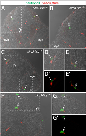
Transverse sections through the head of nlrc3-like–/– embryos at 2.5 dpf show neutrophils that have infiltrated the brain parenchyma and other locations, Related to Figure 4. Cryosections through the head of double transgenic nlrc3-like mutant embryos labeled by neutrophil reporter lyz:EGFP and vasculature marker kdrl:mCherry- CAAX. Images show overlay of GFP and mCherry expression on the bright field image. (A) In nlrc3-like mutants, lyz:EGFP + neutrophils are found intermixed with cells in the brain parenchyma independent of the vasculature in red (arrows). (B) Higher magnification of A as demarcated by the white dotted box. Neutrophils in the mutant are also often found within or surrounding eye vasculature (C–E). (D–E) Higher magnification of the boxed regions in C. (D′–E′) Red and green fluorescent overlay of the same image as D–E respectively. (F–G) Neutrophils are also associated with brain vessels (arrow) and in or near the dorsal head epithelium (arrowhead). |