
Fig. S5
- ID
- ZDB-FIG-140310-7
- Publication
- Thakur et al., 2014 - Dysregulated phosphatidylinositol signaling promotes endoplasmic reticulum stress-mediated intestinal mucosal injury and inflammation in zebrafish
- Other Figures
- All Figure Page
- Back to All Figure Page
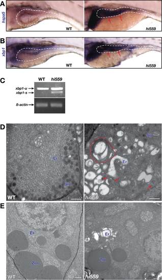
Unresolved ER stress pathology of hi559 GI tissues. (A-B) ISH shows elevated expression of the crucial ER stress-UPR markers hspa5 (A) and xbp1 (B) in the hi559 GI tissue. (C) RT-PCR showing activation of xbp1 by increased transcription of unspliced (xbp1-u) and spliced xbp1 (xbp1-s) in 5-dpf hi559 GI tissues. β-actin was used as RT-PCR control. (D-E) TEM images of 6-dpf wild-type (left panels) and hi559 pancreatic cells (right panels). (D) Ultrastructure of the secretory granulescontaining endocrine pancreatic cells shows increased ER luminal expansion and vacuolization, multi-lamellar autophagic bodies (red outline), and increased mitophagy (red arrow) in hi559. (E) ER luminal expansion is also apparent in the hi559 pancreatic acinar cells containing zymogen granules. Er: endoplasmic reticulum; Sg, secretory granules; Zm; zymogen granules; Mt, mitochondria. Scale bars: D-E, 500 nm. |
| Genes: | |
|---|---|
| Fish: | |
| Anatomical Terms: | |
| Stage: | Day 5 |