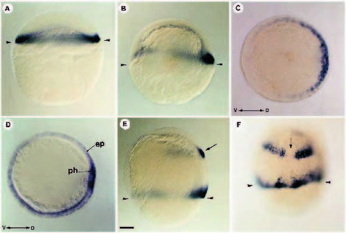
Localization of FGF-8 RNA during blastula and gastrula stages. (A) At blastula stage (30% epiboly), FGF-8 RNA is detected all around the margin of the blastoderm. (B) At early gastrula (50% epiboly) FGF-8 is restricted to the dorsal part of the marginal region. (C) Animal pole view of an embryo at 50% epiboly showing that fgf-8 is expressed as a dorsoventral gradient. (D) At midgastrula stage, FGF-8 transcripts are confined to the epiblast cells (ep) in contact with the underlying hypoblast. Dorsally, paraxial hypoblast (ph) territory is also positive for FGF-8 transcripts. (E) In addition to the marginal gradient, fgf-8 at midgastrula is expressed in the presumptive hindbrain (arrow). (F) Two bands of cells expressing FGF-8 are separated by a nonexpressing domain (arrow) which will later give rise to the ventral midline of the hindbrain. (A,B,E) Lateral views; (C) animal pole view; (D) vegetal pole view; (F) dorsal view. V, ventral; D, dorsal. Arrowheads in A,B,E,F indicate the position of the margin. Scale bar : 100 μm
|

