
Fig. 3
- ID
- ZDB-FIG-140304-36
- Publication
- Zhang et al., 2014 - p35 promotes the differentiation of amacrine cell subtype in the zebrafish retina under the regulation of egr1
- Other Figures
- All Figure Page
- Back to All Figure Page
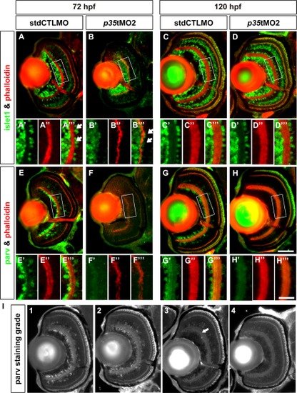
p35 knockdown compromised the differentiation of Parv+ but not Islet1+ ACs. Immunohistochemical analysis of ACs in the controls (stdCTLMO) and p35 morphants (p35tMO2) was performed with anti-Islet1 (Islet1; A–D) and anti-Parvalbumin (Parv; E–H) for cholinergic and a subset of GABAergic ACs, respectively, at 72 and 120 hpf. These markers are shown in green, while the plexiform layers are highlighted by phalloidin in red. A magnified view of the white box is shown at the bottom of each whole retinal section. For Islet1+ ACs, they were not aligned regularly on the boundary of the apical IPL at 72 hpf (A′′′, B′′′; white arrows). In addition, their numbers were also reduced. By 120 hpf, these differentiation problems were largely resolved and the two conditions became comparable (C-C′′′ and D-D′′′), indicating that the effect was caused by a developmental delay. Interestingly, Parv+ ACs were almost absent in the p35-morphant retinas compared with the controls at 72 hpf (E-E′′′ and F-F′′′) and was still substantially reduced at 120 hpf (G-G′′′ and H′-H′′′). The differentiation defects of these ACs likely contributed to the thinning of the IPL at 72 hpf. In addition, the reduction of Parv signal at 120 hpf was quantified as described in the Experimental Procedures section and Table 1. I: An example of the four different staining grades. The white arrow in grade 3 indicates a small region with positive Parv projections detected in the IPL. For A–H: The cryosection of a whole retina is shown at the top and a magnified view of the white box in the central retina is shown at the bottom. For the whole retinal images, lateral is to the left and dorsal is up. Scale bars for the whole retinal section and magnified view were 50 and 25 μm, respectively. |
| Antibody: | |
|---|---|
| Fish: | |
| Knockdown Reagent: | |
| Anatomical Term: | |
| Stage Range: | Protruding-mouth to Day 5 |